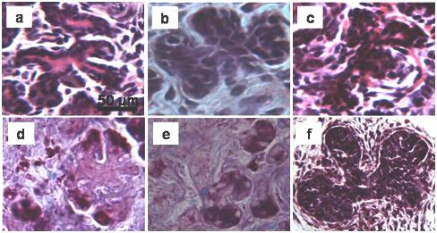
Fig. 3.

Branched structures occurring in the 3 days deligated tissue (a, b, d, e) and in the 2 weeks ligated tissue (c). The AB/PAS staining shows presence of glycoproteins inside the immature acini at the end of the branched structures (d, e). (f) Branched structures occurring during the embryonic development of rat submandibular gland, (embryonic day 18).
