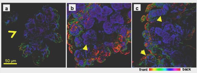
Fig. 5.

Actin immunofluorescence in adult rat submandibular gland. Collagenase-digested cells were incubated with an anti-smooth muscle actin antibody and viewed by confocal microscopy. Optical sections (approximately 1μm) were taken and projected to create a 3D image and colour-coded according to the depth of field. a) Unoperated submandibular gland, star-shaped myoepithelial cells surround the acini, but not the big ducts (open arrowhead). b) In the ligated gland myoepithelial cells have lost their typical morphology but reveal the presence of numerous shrunken acini (arrowhead). c) In the de-ligated gland the myoepithelial cells on the acini recovered their normal star-shaped morphology (arrow). In ligated and deligated tissue myoepithelial cells were found on the big ducts (arrow head).
