
Fig. 7.
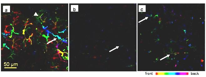
AQP5 immunofluorescence in rat submandibular gland. Collagenase-digested cells were incubated with an anti-AQP5. All images were taken using the same settings; different colours represent differences in depth of field. a) In the control gland AQP5 is strongly expressed along the apical membrane, the intercellular secretory canaliculi (arrowhead) and along the intercalated ducts (arrow). b) In the ligated tissue weak AQP5 expression is restricted to the apical membrane of presumed acinar cells (arrow). c) Some acini in the deligated tissue have regained some AQP5 expression along the intercellular secretory canaliculi (arrow)