
Fig. 8.
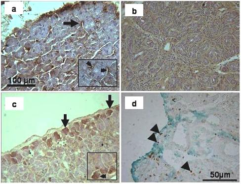
SMG-B immunohistochemistry of the submandibular gland. a) In the unoperated control the staining appeared in some intercalated ducts (arrow; see also double arrow in inset) but not in the acini. b) The 2 weeks ligated gland did not show any localised staining in the parenchyma. c) In the de-ligated tissue, some acini on the edge of lobule showed positive immunoreactivity (arrow; see also double arrow in inset). d) Ki-67 staining (counterstained with Light Green) on 3 day de-ligated plus 2 week ligated submandibular gland. Some cells on the edge of lobules (arrowheads) and in the centre of glands (arrowheads) were labelled.