
Figure 4.
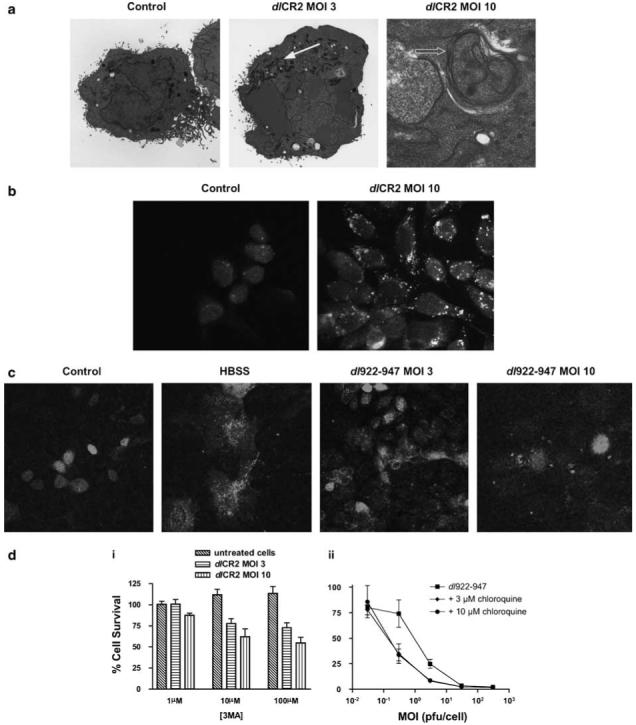
Autophagy. (a) IGROV1 cells were prepared for TEM 72 h following infection with dlCR2 and Ad LM-X (both MOIs 3 and 10). Features of autophagy include increased numbers of lysosomes (closed arrow), the presence of autophagosomes with double membranes (open arrow). (b) IGROV1 cells were infected with dl922-947 (MOI 10) and imaged 48 h later following staining with monodansylcadaverine. (c) IGROV1 cells were co-infected with dl922-947 (MOI 10) and Ad GFP-LC3 (MOI 0.2) and assayed 72 h later for the formation of autophagosomes by confocal microscopy. As positive control, cells infected with Ad GFP-LC3 only were incubated in HBSS for 16 h prior to confocal imaging. (d) (i) IGROV1 cells were infected with dlCR2 (MOI 3 and 10) in the presence of 3-methyladenine (3MA; 0-100 μm). Cell viability was assessed 72 h later. (ii) IGROV1 cells were infected with dl922-947 (MOI 0.03-300) and re-fed with 0-10 μm chloroquine 3 h later. Cell survival was assayed 120 h later and results represent survival normalized for mock-transfected cells at each dose of chloroquine.