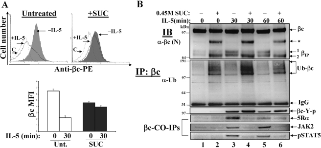
Fig. 7.
Reduction of clathrin-dependent endocytosis alters IL-5-mediated βIP generation and signaling. (A) Cytokine-starved TF1 cells (1×10⁶ cells/tube) were either left untreated (upper left panel) or treated with 0.45 M sucrose (upper right panel) for 30 min to disrupt clathrin lattices. βc cell surface expression was measured by flow cytometry before (–IL-5, shaded histograms) and after 30’ IL-5 stimulation (+IL-5, solid black line). The hatched line represents cells labeled with an isotype-matched control antibody (C). Mean fluorescence intensities (MFI) are plotted and sem are listed in the text. βc: Untreated n = 3; n = 3 for SUC. (B) WCL were prepared from cells treated as described in 7A, IP with anti-βc mAb (S-16), and serially IB with anti-βc, anti-Ub, anti-phosphotyrosine (4G10), anti-IL-5Rα, anti-JAK2, and anti-pSTAT5. Note how the proteolytic processing of βIP is altered, and how ubiquitinated βc receptors accumulate in the presence of sucrose-treated cells (even-numbered lanes). Also, note that CO-IPs of 5Rα, JAK2, and pSTAT5 are blocked in the sucrose-treated lanes.

