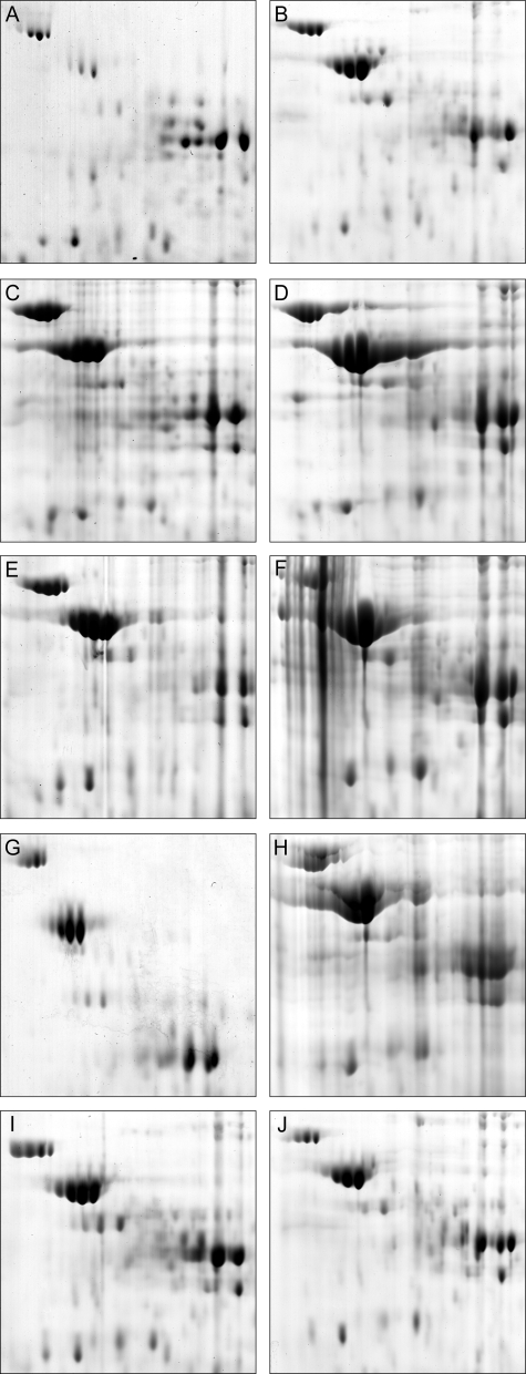
FIG. 7.
Comparison of the proteomes of R. eutropha PHB−4 and various mutants. Sections of 2D PAGE gels (pH ca. 5 to 6; mass, ca. 70 to 35 kDa) loaded with protein extracts from the cells after 64 h and 72 h of cultivation in TSB medium supplemented with 0.5% (wt/vol) sodium gluconate are shown. After withdrawal of the first sample after 64 h of cultivation, NH4Cl was added to a concentration of 0.1% (wt/vol). A second sample was withdrawn after 72 h of cultivation. The sections represent zones in which the flagellin protein occurred when expressed. The strains of R. eutropha were as follows: H16, 64 h (A); H16, 72 h (B); ΔphaP1, 64 h (C); ΔphaP1, 72 h (D); ΔphaP1234, 64 h (E); ΔphaP1234, 72 h (F); PHB−4, 64 h (G); PHB−4, 72 h (H); HF09, 64 h (I); and HF09, 72 h (J).

