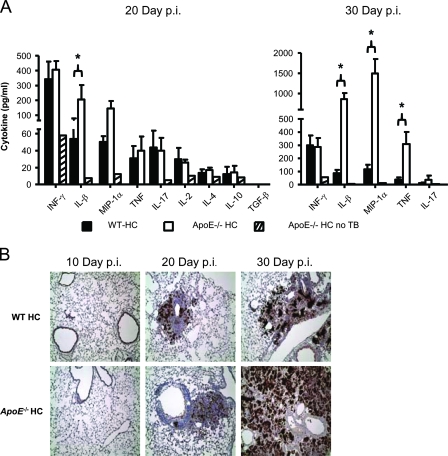
FIG. 4.
Production of inflammatory cytokines in ApoE−/− HC lungs increases with the M. tuberculosis burden, while iNOS production is comparable to that in WT HC lungs. (A) Cytokine production in lung homogenates from WT HC and ApoE−/− HC mice 20 days or 30 days after aerosol infection with M. tuberculosis. The values are the mean cytokine concentrations expressed as pg/ml plus standard deviatons. *, P < 0.05 (n = 4). Lung homogenate from an uninfected ApoE−/− HC mouse was used as a background control. (B) Lung tissue sections from WT HC and ApoE−/− HC mice stained for iNOS production by immunohistochemistry 10 days, 20 days, and 30 days after aerosol infection with M. tuberculosis. Cells expressing iNOS are identified by brown staining; magnification, ×40. The lung sections shown are representative of three mice per mouse strain per time point. The results shown are representative of one experiment.

