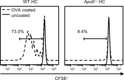
FIG. 6.
In vivo antigen presentation is impaired in ApoE−/− HC mice. Shown are representative histograms of CFSE fluorescence of OT-II CD45.1+ CD4+ cells recovered from the left inguinal LN of WT HC and ApoE−/− HC mice. OT-II cells were stimulated in vivo for 3 days by s.c. injection of OVA-coated or uncoated iron beads near the left inguinal LN. The results are representative of three mice per group that were stimulated with OVA-coated beads; 66.6% and 13.4% of OT-II CD45.1+ CD4+ cells underwent ≥1 cell division in WT HC and ApoE−/− HC mice, respectively. *, P < 0.05 (n = 3). The results shown are representative of one experiment.

