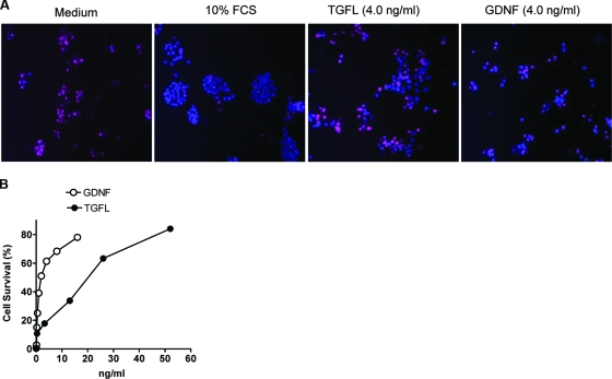
FIG. 6.
TGFL promotes survival of MPC cells. (A) Cells were plated in microtiter wells, cultured overnight in 10% FCS and for 5 days in 10% FCS or 0.5% FCS without (medium) or with TGFL (4 ng/ml) and GDNF (4.0 ng/ml). The microtiter plate was centrifuged at 500 × g for 10 min to increase cell attachment to the substratum. Cell survival was visualized by staining with PI, which stains dead cells red, and Hoechst 33342, which stains live and dead cells blue. (B) TGFL and GDNF promotes cells survival in a dose-dependent manner. The protocol was similar to that for panel A. Cell viability (%) was calculated as 100 × (1 − number of PI-stained cells/total number of cells). Results represent averages of more that 300 cells per point, each in triplicate. This experiment was repeated three times with similar results.

