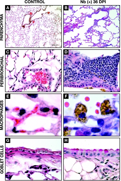
FIG. 1.
Cellular and structural changes in the lungs at 36 days post-N. brasiliensis (Nb) infection. Light microscopy of the lungs from uninfected (control) and N. brasiliensis-infected (day 36 p.i.) BALB/c mice is shown. Histological analysis illustrates focal emphysema-like lesions resulting from parasite migration (A and B) (magnification, ×10; H&E stain), peribronchial infiltration (Panels C & D, 40x magnification, H&E stain), the presence of large alveolar macrophages containing pigmented granules (E and F) (magnification, ×100; H&E stain), and an increase in the number of goblet cells (H) (magnification, ×60; PAS stain).

