FIG. 3.
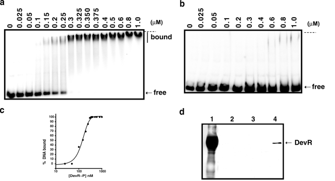
Analysis of DevR binding with narK2-Rv1738 intergenic region. 32P-labeled narK2-Rv1738 intergenic DNA (amplified with narK2F and narK2R primers (Fig. 5a) was incubated with increasing concentrations of DevR ∼P (a) or DevR D54V protein (b). The horizontal dashed line indicates the positions of wells. (c) Fraction of bound DNA (Fig. 3a) versus DevR∼P concentration (see Materials and Methods). (d) Western blot of eluted fractions from DNA pull-down assay developed with anti-DevR polyclonal antibody. Lane 1, purified glutathione S-transferase-tagged DevR protein; lane 2, eluate of reaction with DevR and acetyl phosphate without DNA; lanes 3 and 4, eluate of reactions in which biotinylated narK2-Rv1738 intergenic DNA was incubated with DevR in the absence or presence of acetyl phosphate.

