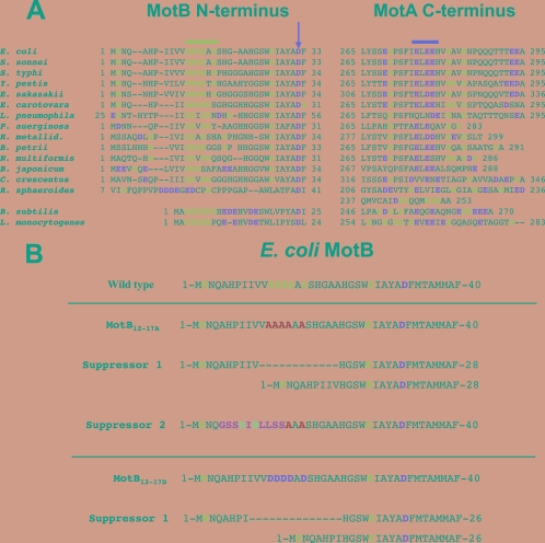
FIG. 1.
(A) Alignment of the N-terminal amino acid sequences of MotB and C-terminal amino acid sequences of MotA for 16 diverse bacterial species. The numbers indicate the positions of the first and last residues in the sequence. Positively charged residues are highlighted blue, and negatively charged residues are highlighted red. The blue and red lines identify the positions of the clusters of positively charged residues in MotB and of negatively charged residues in MotA, respectively. The beginning of the TM helix of MotB is underlined in black, and the absolutely conserved Asp residue that is protonated and deprotonated during proton flow (22) is indicated by the vertical red arrow. The sequences are from Escherichia coli, Shigella sonnei, Salmonella enterica serovar Typhi, Yersinia pestis, Enterobacter sakazaki, Erwinia carotovara, Legionella pneumonophila, Pseudomonas aeruginosa, Ralstonia metallidurans, Bordetella petrii, Nitrosolobus multiformis, Bradyrhizobium japonicum, Caulobacter crescentus, Rhodobacter sphaeroides, Bacillus subtilis, and Listeria monocytogenes. (B) Sequences of the suppressors of the MotB12-17A and MotB12-17D mutations. The top sequence is the first 40 residues of E. coli wild-type MotB. Basic residues are shown in blue, and negatively charged residues are shown in red. The second sequence is for the MotB12-17A mutant. The introduced Ala residues are shown in green. The third and fourth lines are the first suppressor of MotB12-17A (removal of residues 11 to 22). The fifth line is the second suppressor of the MotB12-17A mutant (a +8 frameshift mutation in codon 5 and a −8 frameshift mutation in codons 12 to 14 of motB). Six of the altered residues are shown in orange, whereas the positively charged Arg residues are shown in purple. The sixth line is the MotB12-17D mutant. The introduced Asp residues are shown in red. The seventh and eighth lines show the suppressor of the MotB12-17D mutant (removal of residues 9 to 22).

