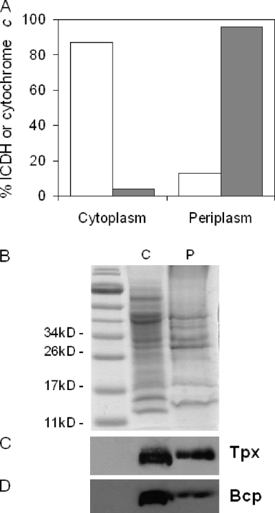
FIG. 8.
Cellular location of Tpx and Bcp proteins in C. jejuni. (A) Distribution of marker proteins in cytoplasm and periplasm after fractionation of wild-type C. jejuni cells according to the method of Sommerlad and Hendrixson (37). Open bars represent the activity of the cytoplasmic enzyme ICDH, and gray bars represent cytochrome c content determined spectroscopically at 550 nm after dithionite reduction (24). (B) Coomassie blue-stained 12% (wt/vol) SDS-polyacrylamide gel showing normalized loadings of cytoplasm (C) and periplasm (P). (C) Immunoblot assay of an identical gel using anti-Tpx antibodies. (D) Immunoblot assay using anti-Bcp antibodies.

