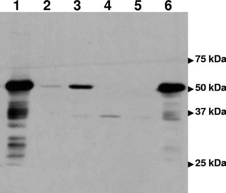
FIG. 3.
Western blotting with FslE antibodies. Whole-cell lysates normalized for cell density were subjected to sodium dodecyl sulfate-polyacrylamide gel electrophoresis, transferred to a polyvinylidene difluoride membrane, and probed with polyclonal antiserum raised to recombinant FslE. The locations of the prestained standards run on the gel are indicated. The lysates are denoted by lanes: 1, GR203 (Δfur) grown in iron-replete CDM; 2, Schu S4 grown in iron-replete CDM; 3, Schu S4 grown in iron-limiting CDM; 4, GR211 (ΔfslE) grown in iron-limiting CDM; 5, vector (pFNLTP6gro-GFP)-transformed GR211 grown in iron-limiting CDM; 6, fslE+ plasmid (pGIR469)-transformed GR211 grown in iron-limiting CDM.

