
Figure 7.
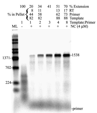
Reverse transcription assay with increasing concentrations of template shows that template, primer, and RT migrate with different fractions depending on the template concentration. Shown is an autoradiogram of an assay using 1.9 kb RNA as template with no excess template (1:1 template:primer) and with increasing concentrations of template (2, 3, 4, and 8 template:primer). Lanes with – indicate reactions without NC and those with +, with NC (4 μM). The percentages of RT activity, primer and template in the pellet at different concentrations of template are indicated and were determined as described in Materials and Methods (not calculated for 3:1 template: primer). Other markings are as in Figure 1(b). A repeat experiment yielded similar results.