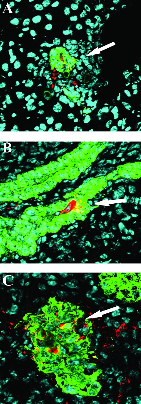
FIG. 7.
Detection of RRV-infected cells in extraintestinal organs using immunofluorescent staining and confocal microscopy. The liver (A), bile duct (B), and pancreas (C) from RRV-infected IFN-αγR KO mice were collected on day 8 p.i. and frozen in OCT. Liver and pancreas sections were costained with anti-VP6 (red) and anti-keratin 8 (green). Bile duct sections were costained with anti-VP6 (red) and anti-keratin 19 (green). Cell nuclei were stained with TOTO-3 iodide (blue). Sections were observed under a confocal microscope. Magnification, ×400. The colocalization of viral antigen and epithelial cell markers is indicated by arrows.

