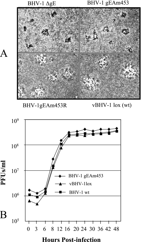
FIG. 4.
(A) Plaque morphology of BHV-1gEΔ, vBHV-1gEAm453, vBHV-1gEAm453R, and vBHV-1lox in MDBK cell monolayers. Viruses were inoculated onto MDBK cell monolayers, overlaid with 1.6% carboxymethyl cellulose, fixed at 48 h postinfection, and stained with 0.35% crystal violet solution. (B) One-step growth curve of BHV-1gEAm453, BHV-1lox, and wt BHV-1 viruses in MDBK cells. Confluent MDBK cells were infected at a multiplicity of infection of 5 PFU per cell with viruses. After 1 h of adsorption at 4°C, residual input viruses were removed. The cultures were washed three times with phosphate-buffered saline, and 5 ml of medium was added to each flask before further incubation (37°C). At the indicated time intervals, replicate cultures were frozen. Virus yields were determined by titration on MDBK cells. Each data point represents the average for duplicate samples obtained from separate infections.

