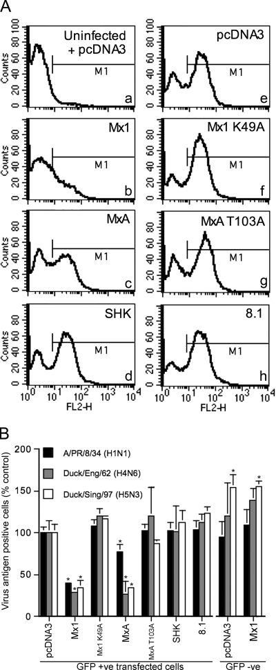
FIG. 2.
Flow cytometric detection of influenza vRNPs in transfected 293T cells. 293T cells were cotransfected with Mx-expressing plasmids (or pcDNA3) (1.5 μg) and pEGFP-C1 (0.5 μg) and infected after 48 h with A/PR/8/34, A/Duck/England/62, or A/Duck/Singapore/97. Fourteen hours postinfection, the cells were stained for vRNP and analyzed by flow cytometry. Panel A shows the level of A/PR/8/34 antigen in GFP-positive cells that had been cotransfected with the indicated plasmids. Panel “a” shows the background staining detected in uninfected, pcDNA3-transfected cells and was used to set the fluorescence threshold marker (M1), which demarcates between antigen-positive and -negative cells. In panel B, the data are derived from independent experiments (A/PR/8/34, n = 5; A/Duck/England/62, n = 3; A/Duck/Singapore/97, n = 3) and the bar heights represent the percentages of antigen-positive cells expressed relative to that for the pcDNA3 control group for each virus. The means (and standard deviations) are shown for either GFP-positive or GFP-negative cell populations from wells that had been transfected with the constructs as shown. *, P of <0.01 relative to GFP-positive pcDNA3-transfected cells.

