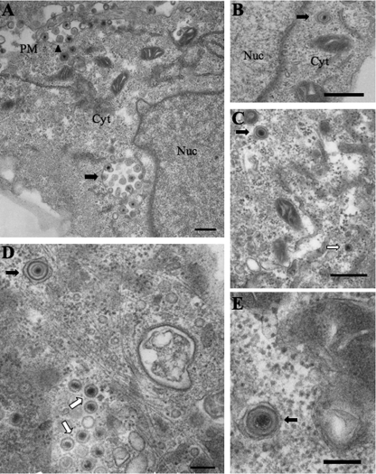
FIG. 6.
Enveloped K26GFP and KΔUL36GFP particles are present in the cytoplasm of infected Vero cells. Enveloped HSV-1 particles were present in the cytoplasm of both K26GFP-infected (A and B) and KΔUL36GFP-infected (C to E) Vero cells. (A) Cytoplasmic enveloped K26GFP particles can be observed in the cytoplasm (black arrow), with a number of enveloped capsids contained within one organelle. Extracellular capsids are also present (black arrowhead), attached to the plasma membrane. (B) An enveloped K26GFP particle is present in the cytoplasm. (C to E) Enveloped KΔUL36GFP particles are present in the cytoplasm (black arrows), and nonenveloped capsids (C and D) were also observed (white arrows). Bars, 500 nm (A to C); 200 nm (D and E).

