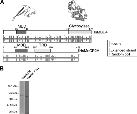
FIG. 2.
MBD4 and MeCP2, two methylated-DNA-binding proteins with different structures. (A) The box drawings are a schematic representation of the functional domain organization of human MeCP2 and MBD4. The secondary structures predicted from the amino acid sequences by using the hierarchical neural network (23) are shown underneath. The tertiary-structure organizations of the methyl-CpG-binding and glycosylase domains of human MBD4 as predicted from the crystallographic data on the methyl-CpG-binding domain (MBD) of human MBD1 (50) and the mouse glycosylase domain of MBD4 (71) are shown above the corresponding schematic linear representations. Tertiary-structure predictions were carried out using the SWISS-MODEL server (57). TRD, transcription repression domain; Hs, Homo sapeins. (B) SDS-PAGE analysis of the recombinant MBD4 and MeCP2 forms used in this work. The numbers on the left-hand side of the gel correspond to the molecular masses of the proteins as indicated by a marker.

