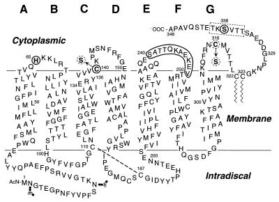
Figure 1.
A secondary structure model of bovine rhodopsin shows the single and double Cys replacements in the cytoplasmic domain made in this work. The amino acids in the C-terminal peptide chain shown boxed (dotted rectangle) were replaced one at a time by Cys residues to prepare single Cys containing mutants. In every case, the double Cys mutants contained the first Cys at position 338 and the second Cys at position 65 in AB loop; position 140 in CD loop is shown by solid circles or one of the positions in EF loop is shown by the solid boundary. The native Cys at positions 140 and 316 were replaced by serines (dotted circles) in all the single and double Cys mutants except for C140/S338C, which contains the Cys at position 140.

