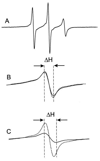
Figure 5.
The EPR spectra of spin labeled rhodopsin mutants. (A) the EPR spectrum of S338R1 in a solution of DM in the dark state. The scan width in the magnetic field is 100 G; (B) the center lines (m1 = 0) for the sum of the S338R1 and T243R1 single mutants (dashed line) and the T243R1/S338R1 double mutant (solid line); (C) the center lines for the sum of the S338R1 and K245R1 single mutants (dashed line) and the K245R1/S338R1 double mutant (solid line). In B and C, the magnetic field scan width is 17 G. ΔH is the peak-to-peak width of the center line of the corresponding double mutant.

