Figure 1.
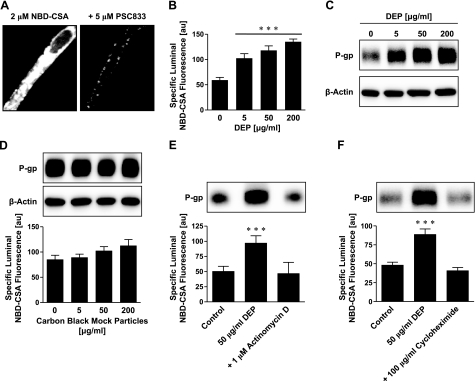
DEPs increase P-glycoprotein expression and transport activity in isolated rat brain capillaries. A) Left panel: representative image of an isolated brain capillary after 1 h of exposure to 2 μM NBD-CSA, showing steady-state NBD-CSA fluorescence. Note that NBD-CSA transport is concentrative from bath (no visible fluorescence) to endothelium (low fluorescence) to capillary lumen (high fluorescence). Right panel: the P-glycoprotein-specific inhibitor PSC833 blocks concentrative NBD-CSA transport from capillary endothelium to capillary lumen. B) DEPs increase specific luminal NBD-CSA fluorescence after 6 h in a concentration-dependent manner. C) Western blot analysis showing concentration-dependent P-glycoprotein up-regulation through DEPs in brain capillary plasma membranes. β-Actin was used as loading control. D) CB mock particles had no effect on P-glycoprotein expression or transport activity after 6 h. P-glycoprotein transport activity is shown as specific luminal NBD-CSA fluorescence. E) Inhibition of transcription with actinomycin D abolishes DEP-induced increase in P-glycoprotein expression (Western blot) and transport activity (specific luminal NBD-CSA fluorescence). F) Inhibition of protein synthesis with cycloheximide also abolishes DEP up-regulation of P-glycoprotein. For Specific luminal NBD-CSA fluorescence values are means ± se for 10 capillaries from a single preparation (pooled tissue from 10 rats); arbitrary units (au; scale 0–255). ***P < 0.001 vs. control.

