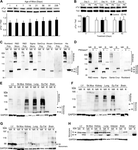
Figure 2.
Tie2 activation is stable during cardiac remodeling and heart has abundant ang1 monomers compared to other tissues. A, B) Tie2 phosphorylation [immunoprecipitation and Western blotting for phosphotyrosine (pTyr) and tie2] shows no change during physiological (A) and pathological (PE-induced) (B) LV remodeling, remaining stable in neonatal through adult (A) and control vs. hypertrophic LVs (B). Studies done in triplicate; n = 3/group. C, D) Results of Western blotting conducted with rhAng1 (0.09 μg) in nonreduced (NR) and reduced (R) (βMe) conditions. Blots were probed with anti-ang1 monoclonal (mono) or polyclonal (poly) antibodies in duplicate studies. The ang1 monomers and multimers detected vary (C). PNGase-F treatment of rhAng1 reveals that some ang1 forms were glycosylated (D). E–H) Western blot analysis of protein lysates from mouse (E–G) and human (H) LV, skeletal muscle (sk. mus), kidney, lung, epididymal fat (ep. fat), and brain tissue, evaluated in nonreduced and reduced (βMe) conditions. Blots were probed with anti-ang1 [monoclonal: R&D (E) and Sigma (F); polyclonal: Rockland (G, H)] and anti-GAPDH antibodies in duplicate studies. Ang1 monomers and multimers are seen in mouse (E–G) and human (H) adult tissues. Heart and skeletal muscle show low multimer levels (E, F) and high monomer levels (G) vs. other tissues.

