Figure 1.
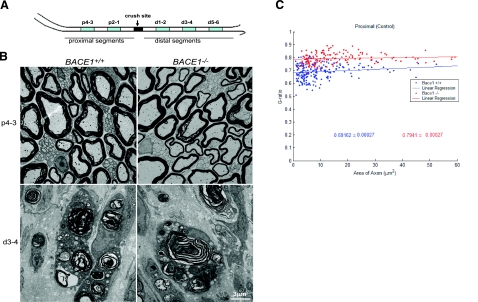
Sciatic nerve crush causes Wallerian degeneration. A) Sciatic nerves of 2-month-old mice were crushed at the midthigh. Blue segments (1 mm in length) from the distal and proximal ends were examined by electron microscopy. Numbers indicate distance from the crush site. B) Ultrastructure of proximal segments p4–3 and distal segments d3–4 in WT and Bace1-null mouse nerves at 5 days after crush. As in uncrushed nerves, proximal nerve segments are hypomyelinated in BACE1-null mice. In the distal segments, axons have degenerated, and myelin debris is abundant in macrophages in both nerves. Scale bar = 3 μm. C) Scatter plot of g ratio of myelinated fibers in the proximal stump 5 days after crush. Each spot represents the g ratio of one myelinated fiber. The g ratio was calculated by dividing the inner circumference of the axon (without myelin) by the outer circumference of the total fiber (including myelin). Data are presented as mean ± se. Blue: WT mice; red: BACE1-null mice; n = 3; P < 0.001.

