Figure 1.
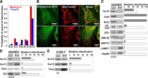
Endogenous Gα12 is localized to mitochondria in HUVECs and COS-7 cells. A) Prediction of mitochondrial targeting of different Gα subunits using Mitoprot II (red) and TargetP (blue). UniProt accession numbers: Gαs, P63092; Gαi1, P63096; Gαq, P50148; Gα12, Q03113; Gα13, Q14344. B) Colocalization of endogenous Gα12 with MitoTracker in HUVECs. Living cells were incubated with MitoTracker, fixed, and immunostained with Gα12-specific antibodies (top and bottom panels: lots H052 and I150, respectively). Scale bars = 20 μm. C) Distribution of Gα12 and organelle markers in the mitochondrial fraction (P10) and the fraction containing other organelles except nuclei (S10). P10 was resuspended in 1/12.5 of initial volume, and equal volumes of P10 and S10 were loaded on the gel. The following marker proteins were examined: COX and Bcl-2 (mitochondria), VE-cadherin, and zonula occludens-1 (ZO1) (plasma membrane/microsomal fraction), GM130 (Golgi apparatus), GRP72 (endoplasmic reticulum), LAMP1 (lysosomes), as well as Hsp90 as a representative of proteins mainly present in the cytoplasm. D, E) Distribution of subunits of different heterotrimeric G proteins in HUVECs and in COS-7 cells, respectively. In E, P10 was resuspended in 1/4 of initial volume. Experiments shown were repeated twice with similar results.

