Figure 2.
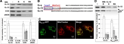
N terminus of Gα12 targets proteins to the mitochondrial surface. A) Mitochondrial fraction of HUVECs was subjected to hypotonic rupture (R) and/or treated with Proteinase K (K) as indicated. The effect of these treatments on Gα12, Gα13, COX, and eNOS was assessed by Western blotting with corresponding antibodies. Data were normalized to the protein content in untreated aliquots. Data shown are the means of two replicates; error bars show values obtained in each replicate. B) Sequences in Gα12 predicted by Mitoprot II (red) and TargetP (blue) to function as signal peptide for mitochondrial targeting. UniProt accession numbers: Gαq, P21279; Gα12, P27600; Gα13, P27601. C) Mitochondrial targeting of NGα12-GFP (GFP fused to the N terminus of Gα12, underlined in B). Twenty-four hours after transfection, living HUVECs were incubated with MitoTracker and examined using confocal microscopy. D) N terminus of Gα12 does not mediate GFP translocation into mitochondrial matrix. Mitochondrial fraction obtained from COS-7 cells (either nontransfected or transfected with NGα12-GFP) was subjected to Proteinase K protection assay as in A. Endogenous Gα12 and NGα12-GFP were detected by Western blotting with Gα12-specific antibody. Data are means of 3 replicates; error bars indicate sd.

