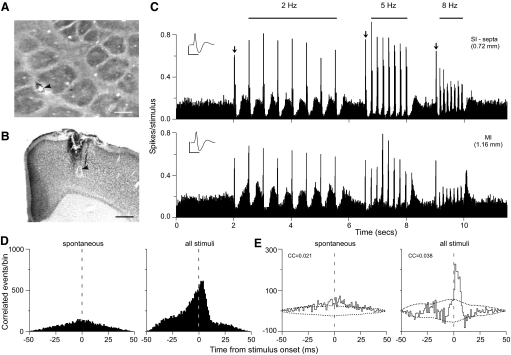
FIG. 9.
Example of coordination between a septal neuron and an MI neuron shown as in Fig. 8. A: recording site in the SI septa. Scale: 250 μm. B: recording site in MI cortex. Scale: 500 μm. C: PSTHs of the SI and MI neuronal responses. Bin widths: 10 ms; waveform scales: 1 mV, 1 ms. D: raw CCGs show spontaneous and stimulus-induced coordination of SI septal neuron and the MI neuron. E: shift-corrected CCGs for the same neuron pair. Bin widths: 1 ms.

