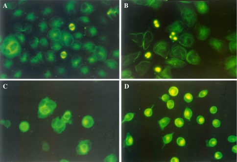
Fig. 7.
Immunofluorescence images of CNE-2Z cells treated with TBMS1. Cells were cultured for 24 h and then incubated without (a) or with 2.5 μM paclitaxel (b), 2.5 μM colchicine (c) and 25 μM TBMS1 (d) for 3 h. The fixed cells were stained with the anti-tubulin primary antibody and the fluorescein 5-isothiocyanate-conjugated secondary antibody as described in “Materials and methods”. Magnification ×400

