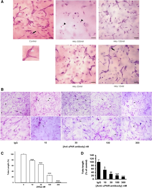
Fig. 1.
Effect of two-chain high-molecular-weight kininogen (HKa) and anti-urokinase plasminogen activator (uPA) receptor (uPAR) antibody on human umbilical vein endothelial cell (HUVEC) morphogenesis in three-dimensional (3-D) collagen-fibrinogen gel (22-h incubation). A: inhibition of capillary tube formation by HKa at different concentrations. HUVECs were cultured in 3-D collagen-fibrinogen gel matrixes with or without HKa for 22 h at 37°C. HUVECs were then fixed with 4% paraformaldehyde and stained with 1% toluidine blue. Black arrows point to vacuoles. A vacuole is an open space within a cell or between 2 cells only. Black rrowheads point to apoptotic cells. The lumen to the white arrow pointed to is magnified below. A lumen is a space formed by 3 or more cells. Bars = 100 μm. Magnification: ×200. B: inhibition of tube formation by mouse monoclonal anti-uPAR antibody at different concentrations. Concentrations of the monoclonal antibodies (in nM) were calculated by measuring the protein concentration of the antibodies and dividing them by molecular weight (150 kDa). HUVECs were cultured in 3-D collagen-fibrinogen gel matrixes with or without anti-uPAR antibody for 22 h at 37°C. HUVECs were then fixed with 4% paraformaldehyde and stained with 1% toluidine blue. Black arrows point to intracellular vacuoles. White arrows point to the lumen. Magnification: ×100 (top) and ×200 (bottom). C and D: quantification of tube length formation after an incubation with HKa (C) or anti-uPAR antibody (D). A tube is a cellular structure that measures >100 μm in length and is visibly formed by the body of 3 cells or more. Tube length was measured in μm/mm2. Results were normalized and reported as percentages of the tube length (means ± SE). All treatments were compared with the control. *P < 0.05; **P < 0.01; ***P < 0.005.

