Fig. 5.
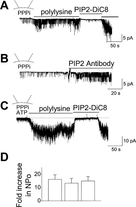
Activation of TRPA1 by polylysine and PIP2 antibody in inside-out patches. A and B: inside-out patch was formed in the presence of 5 mM PPPi in the bath solution. Addition of polylysine (10 μg/ml; A) or PIP2 antibody (20 μg/ml; B) caused activation of TRPA1. Subsequent addition of PIP2 (10 μM) quickly and reversibly inhibited the channels. C: inside-out patch was formed in the presence of 5 mM PPPi and 2 mM ATP in the bath solution. Addition of polylysine (10 μg/ml) caused activation of TRPA1 and further addition of PIP2 (10 μM) inhibited the channels. D: bar graph summarizes the data from A–C. Each bar is the mean ± SD of 5 determinations. No significant differences were present (P > 0.05).

