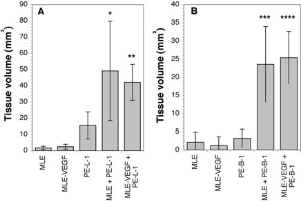
Figure 2.
Growth of prostatic cells under the renal capsule. MLE-ßgal mouse endothelial cells (2.5 × 10⁵ cells) transfected with an expression vector encoding VEGFA (MLE-VEGF) or with the vector alone (MLE) were co-inoculated under the renal capsule with 5 × 10⁵ PE-L-1 prostate luminal epithelial cells (A) or PE-B-1 prostate basal epithelial cells (B). As a control 7.5 × 10⁵ MLE, MLE-VEGF, PE-L-1, or PE-B-1 cells were implanted alone. After 60 days, the mice were sacrificed, and the size of the prostatic growth measured. * p < 0.01 compared to MLE alone and p < 0.05 compared to PE-L-1 alone; ** p < 0.01 compared to MLE-VEGF alone or PE-L-1 alone; *** p < 0.01 compared to MLE alone or PE-B-1 alone; **** p < 0.01 compared to MLE-VEGF alone or PE-B-1 alone.

