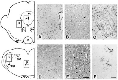
Figure 1.
Neuropathology of Tg(BoPrP)Prnp0/0 mice inoculated with BSE prions. (A) No pathological changes were found in the periaqueductal grey of the midbrain. (B) Mild to moderate vacuolar degeneration was found in the reticular formation of the midbrain tegmentum. (C) Reactive astrocytic gliosis colocalized with sites of vacuolar degeneration: astrogliosis in the red nucleus is shown here. (D) Little or no vacuolar degeneration was found in the tract or the nucleus of the spinal tract of the trigeminal nerve in the medulla. (E) Moderate to severe vacuolar degeneration occurred in the medial tegmentum of the medullary reticular formation. (F) Small PrP-immunopositive primitive plaque-like deposits colocalized with sites of the most severe vacuolar degeneration. Diagrams show locations of photomicrographs. Hematoxylin and eosin stain was used in A, B, D, and E. Glial fibrillary acidic protein immunohistochemistry was used in C. PrP immunohistochemistry was used in F. Bar in E = 100 μm and also applies to A, B, and D. Bar in F = 50 μm and also applies to C. Italicized letters identify selected brainstem structures: CP, cerebral peduncle; IP, interpeduncular nucleus; PG, periaqueductal grey; Py, pyramidal tract; RN, red nucleus; Sol, nucleus and tractus solitarius; Sp5, nucleus of the spinal tract of the trigeminal nerve.

