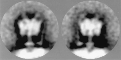
Figure 2.
Side view projections of the V-ATPase of C. fervidus incorporated in liposomes. Shown are two out of six classes of a subgroup of 240 side view projections of the V-type ATPase of C. fervidus incorporated in liposomes. They show V1 in a trilobed view and, besides a central stalk, a peripheral second stalk in two positions. For the left and right images, 39 and 42 images were summed, respectively. The diameter of the area shown is 320 Å.

