Fig. 5.
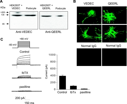
Differentiated cells from a podocyte cell line express functional BKCa channels. A: immunoblot analysis using isoform-specific antibodies showing expression of Slo1VEDEC and Slo1QEERL in differentiated cells of a podocyte cell line. Adjacent lines are extracts of HEK-293T cells expressing the corresponding Slo1 isoform. The antibodies used in this analysis were described previously and do not cross-react (16). B: confocal analysis using the same antibodies also shows that both isoforms were expressed in differentiated podocytes. Note signal in paranuclear intracellular compartments and in foot processes. Control images are from cells in which rabbit IgG was used as primary antibody. C: endogenous BKCa currents in podocytes revealed by whole cell recordings using microelectrodes containing 5 μM free Ca2+. Note very slow activation kinetics and partial blockade after treatment with 500 nM iberiotoxin (IbTX) and complete blockade after 1 μM paxilline. Bar graph at right shows means ± SE for repetitions of this experiment (n > 10 cells per group). Mean currents in all 3 groups are significantly (P < 0.05) different from each other, as determined using one-way ANOVA and Tukey's honest significant difference test for unequal sample size.

