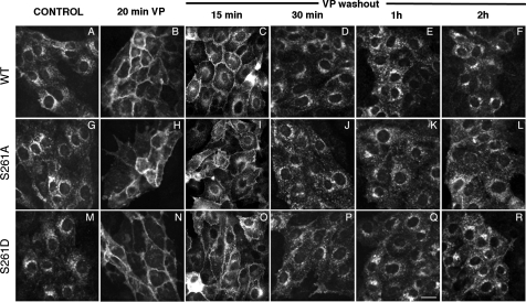
Fig. 4.
Distribution of wild-type AQP2, AQP2-S261A, and AQP-S261D during VP treatment and washout. Both AQP2-S261A, AQP2-S261D, and wild-type AQP2 (AQP2-WT) were present in the perinuclear region in the cytoplasm under control, nonstimulated conditions (A, G, and M). Upon treatment with VP for 20 min, AQP2-S261A, AQP-S261D, and AQP2-WT accumulated on the plasma membrane (B, H, and N). After washout of VP for 15 min, plasma membrane staining of AQP2 wild-type, S261A, and S261D remains but intracellular staining for AQP2 is more abundant than with no VP washout (compare B, H, and N with C, I, and O). After washout for 30 min, plasma membrane staining of AQP2 wild-type, S261A, and S261D was reduced, reflecting AQP2 internalization (D, J, and P). Between 1 and 2 h after washout, AQP2 wild-type, S261A, and S261D were located mainly in the perinuclear region of the cytoplasm (E, K, Q, F, L, and R). There was no obvious difference in the rate at which the plasma membrane staining of AQP2 was diminished over time after washout in these 3 cell lines. Bar = 20 μM.

