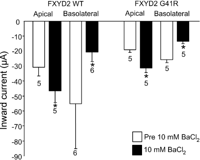
Fig. 8.
Transepithelial current measurements in FXYD2 WT or G41R-expressing MDCK cells. The transepithelial currents at −80 mV from WT and G41R MDCK cells when exposed to a large transepithelial Mg2+ gradient and in the absence (open bars) or presence (filled bars) of 10 mM Ba2+ at the apical or basolateral surface. *Values that are significantly different from the values in the absence of Ba2+ (P < 0.05).

