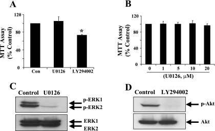
Fig. 4.
Effect of ERK1/2 and PI3K pathway inhibitors on RPTC proliferation. RPTC were cultured in the presence of serum for 24 h and then incubated for an additional 48 h with 10 μM U0126 or 20 μM LY294002 (A), or various concentrations of U0126 as indicated (B). Cell proliferation was determined using the MTT assay. Data are expressed as means ± SD of the percentage of MTT activity compared with controls grown with diluent (n = 3). *P < 0.05, compared with control group. B: RPTC were cultured for 48 h and then treated with 10 μM U0126 or 20 μM LY294002 for 30 min. Cell lysates were prepared and subjected to immunoblot analysis using antibodies to phospho-ERK1/2, phospho-Akt, total ERK1/2, or total Akt. Representative immunoblots from 3 or more experiments are shown.

