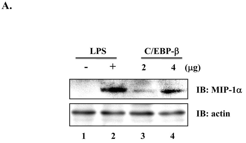
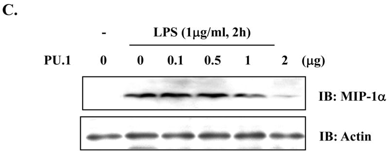
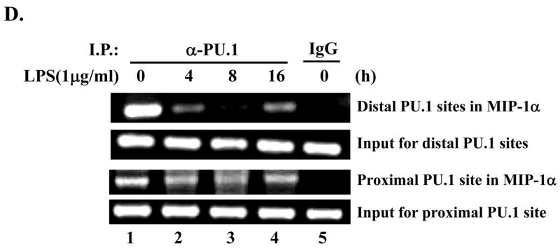
Fig. 4. PU.1 negatively regulates MIP-1α expression.


(A) RAW 264.7 cells were transfected with pcDNA3.1 (lanes 1 and 2) or a plasmid encoding C/EBP-β (lanes 3 and 4) for 48h. Transfection was normalized with pcDNA3.1 to 4μg. Total cell lysate was analyzed by Western blot for MIP-1α (top panel) and actin (bottom panel). (B) MIP-1α expression in PU 5.7 following LPS treatment was determined by Western blot analysis. (C) RAW 264.7 cells were transfected with increasing amounts of a PU.1-expressing plasmid, and the transfected cells were treated with LPS for 2h. MIP-1α expression was determined by Western blot analysis. (D) PU.1 binding to the endogenous MIP-1α promoter was analyzed by ChIP assay. PU.1 bound to DNA was immunoprecipitated by α-PU.1 antibody (lanes 1 to 4), and co-precipitated DNA was analyzed by PCR for distal PU.1 sites (top two panels) and proximal site (bottom two panels). Included was isotypic IgG to exclude a nonspecific immunoprecipitation (lane 5).
