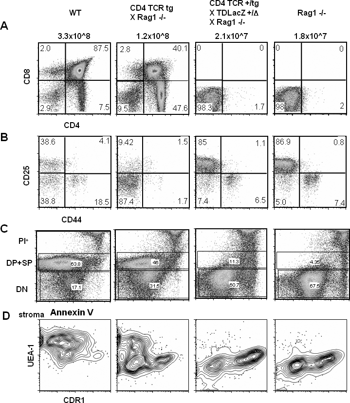
Figure 7. Anti-LacZ–Specific TCR-Bearing Transgenic Thymocytes Are Deleted at the DN Stage in the Presence of TSCOT-LacZ Expression.

(A) CD4 and CD8 thymocyte profiles. The names of each of the four mouse strains used and their total thymocyte yields are shown above the plots. The percentages of the single and double staining subsets are shown in the four quadrants.
(B) CD25 and CD44 profiles of the DN thymocytes with percentages in the quadrants.
(C) Annexin V staining of all cells stained with CD4, CD8, and PI shown in FL3 channel. PI+ cells are at the top, DP and SP cells in the middle, and DN cells in the bottom.
(D) The UEA-1 and CDR1 profiles of the CD45− stromal cell compartment from the four different mouse lines. The genotypes of each mice are verified for the loci of Rag1, TDL, and TCR α and β chains by PCR
