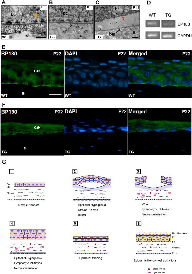
Figure 5. Defective hemidesmosomes and decreased BP180 expression in K14-DN-Clim mice.
(A-C) Electron microscopy ultrastructure of the epithelial-basement region, including hemidesmosomes, in corneas from mice of the indicated genotypes. Red arrow indicates a hemidesmosome on the basal surface of a basal epithelial cell that has separated from the underlying basal lamina. (D) Semiquantitative RT-PCR analysis of BP180 mRNA levels in cornea from wild type (WT) and K14-DN-Clim (TG) mice. GAPDH is included as a control. (E, F) Immunofluorescence analysis of BP180 expression in wild type and K14-DN-Clim corneal epithelium. DAPI staining shows nuclei in the same section. (G) Model for the progression of corneal abnormalities in K14-DN-Clim mice. bc, basal epithelial cell; bm (or BM), basement membrane; ce, corneal epithelium; Endo, endothelium; Epi, epithelium; s, stroma; Scale bar: A, 200nm; C, 500nm; E, 20μm.

