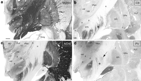
Fig. 11.
Photomicrographs of adjacent frontal sections 15 mm anterior to the posterior commissure stained for a myelin, c AchE, and b immunostained for the calcium-binding proteins CB and d PV. Pallidothalamic fibres (fl, ft) are darkly stained for myelin, moderately for AChE and PV, and negatively for CB. The STh, thalamic VLp nucleus and the optic tract (ot) are strongly enhanced in PV-ir. The arrow in d points to presumed pallidosubthalamic fibres (fasciculus subthalamicus) crossing the posterior limb of the internal capsule. The asterisks in a, b, and d indicate matching locations in the internal capsule corresponding to striatonigral fibres which are strongly enhanced in CB-ir but unstained for myelin. Case Hb4. Scale bar (a): 2 mm

