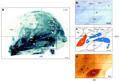
Fig. 3.
Representation of the cerebellothalamic (fct) and pallidothalamic (al, fl, ft) tracts on sagittal section of the atlas (panel c) as delimited from myelin (area comprised in the rectangle in a). Thalamic nuclei and STh were best identified on adjacent Nissl section (panel b). Panel d shows PV immunostaining at same sagittal level. The arrows in a and d point to a small gap separating the fct and ft, visible in myelin and PV-ir, respectively. Notice some PV-ir enhanced fibres in the internal capsule near the anterior pole of the STh and presumably corresponding to the fasciculus subthalamicus. Scale bars (a and c): 2 mm

