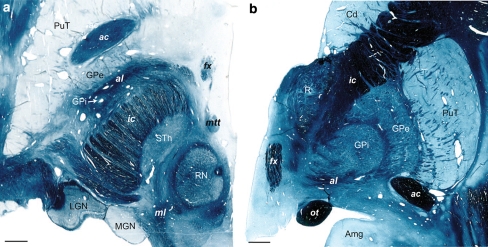
Fig. 9.
a Illustration of the pallidal emergence of al in horizontal and b frontal sections stained for myelin. The levels are 4.5 mm ventral to the intercommissural plane (in a) and 22 mm anterior to pc (in b). Note the relatively large anteroposterior extent of the al at the ventral limit of the GPi in a. Cases Hb5 (left panel) and Hb4 (right panel). Scale bars: 3 mm

