Fig. 4.
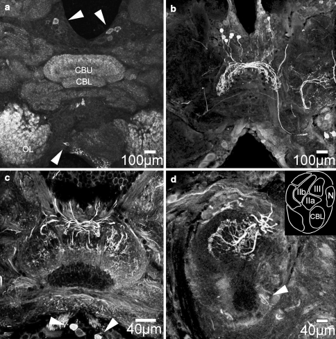
Expression of nitric oxide synthase (NOS) and accumulation of citrulline in the brain of female C. biguttulus. a Horizontal brain section processed for anti-universal NOS immunocytochemistry containing labeled cell bodies in the pars intercerebralis region and the ventro-median protocerebrum (arrowheads) and diffuse staining in the upper division of the central body (CBU) and the glomeruli of antennal lobes (OL). b, c Horizontal brain sections processed for anti-citrulline immunocytochemistry resulted in strong labeling of cell bodies in the pars intercerebralis region and their projections into the upper division of the central body and the lateral accessory lobes. Immunopositive primary neurites either remained ipsilaterally or crossed through the posterior chiasm to the contralateral side before entering the central body. These neurons represent columnar neurons of the central body. Arrowheads in c indicate intensely labeled cell bodies in the ventro-median protocerebrum whose neurites enter the central body through the posterior groove (arrowhead in d) between the lower division (CBL) and the noduli (N). d Parasagittal section through the central body and schematic map of transected neuropils. Citrulline immunolabeled neurites innervate layers II and III of the central body upper division. The lower division of the central body is entirely devoid of citrulline-associated immunolabeling (also visible in c)

