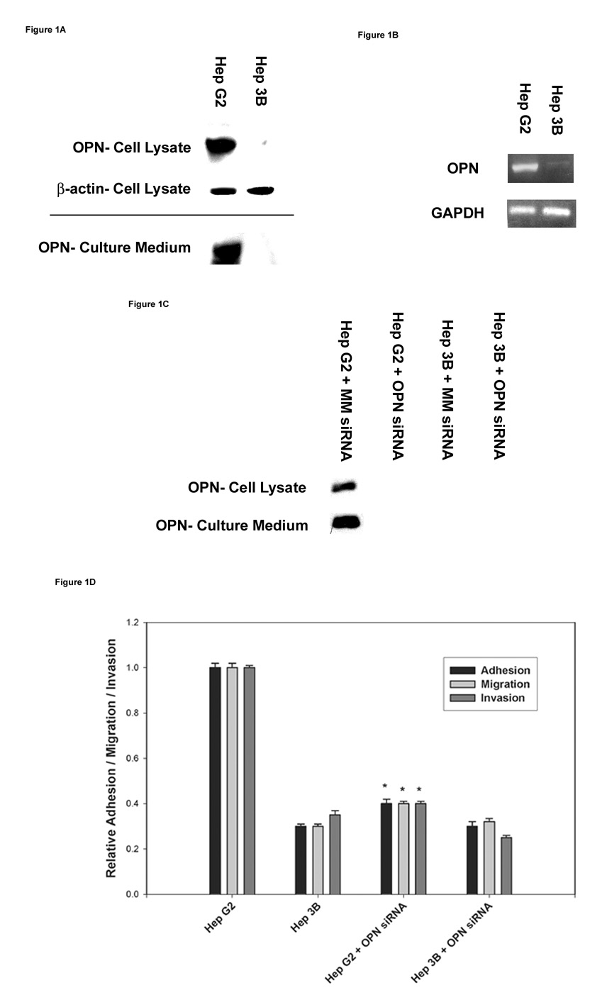
Figure 1. Detection of OPN mRNA and protein expression in the two HCC cell lines.
(A) Total cellular lysates and concentrated culture media from HepG2 and Hep3B cell lines were subjected to immunoblot analysis to determine OPN protein expression.
(B) RT-PCR analysis of OPN mRNA expression in HepG2 and Hep3B cell lines. GAPDH mRNA expression was used as a house keeping control gene.
(C) In vitro suppression of OPN expression by OPN siRNA transfection in the two HCC cell lines. HepG2 and Hep3B cell lines were transiently transfected with either MM siRNA (Mismatch siRNA) or OPN siRNA for 48 h. OPN protein expression was determined by western blot analysis. Blots (A–C) are representative of three separate experiments.
(D) Functional properties of the two HCC cell lines were assessed by their adhesion, migration and invasion characteristics as described in materials and methods. Correlation of OPN expression to its metastatic behavior in HepG2 and Hep3B cells was determined by specifically inhibiting OPN expression by standard OPN siRNA techniques. Data are expressed as mean ± SEM (* p< 0.05 vs. HepG2).

