
Figure 2. Western blot detection of AQP2 in plasma membrane-enriched fractions from LLC-PK1 cells expressing c-myc-tagged AQP2 (A). Indirect immunofluorescence microscopy of tissue slices showing AQP2 redistribution in the inner stripe (outer medulla) of collecting duct principal cells in response to PDE V inhibition (B).
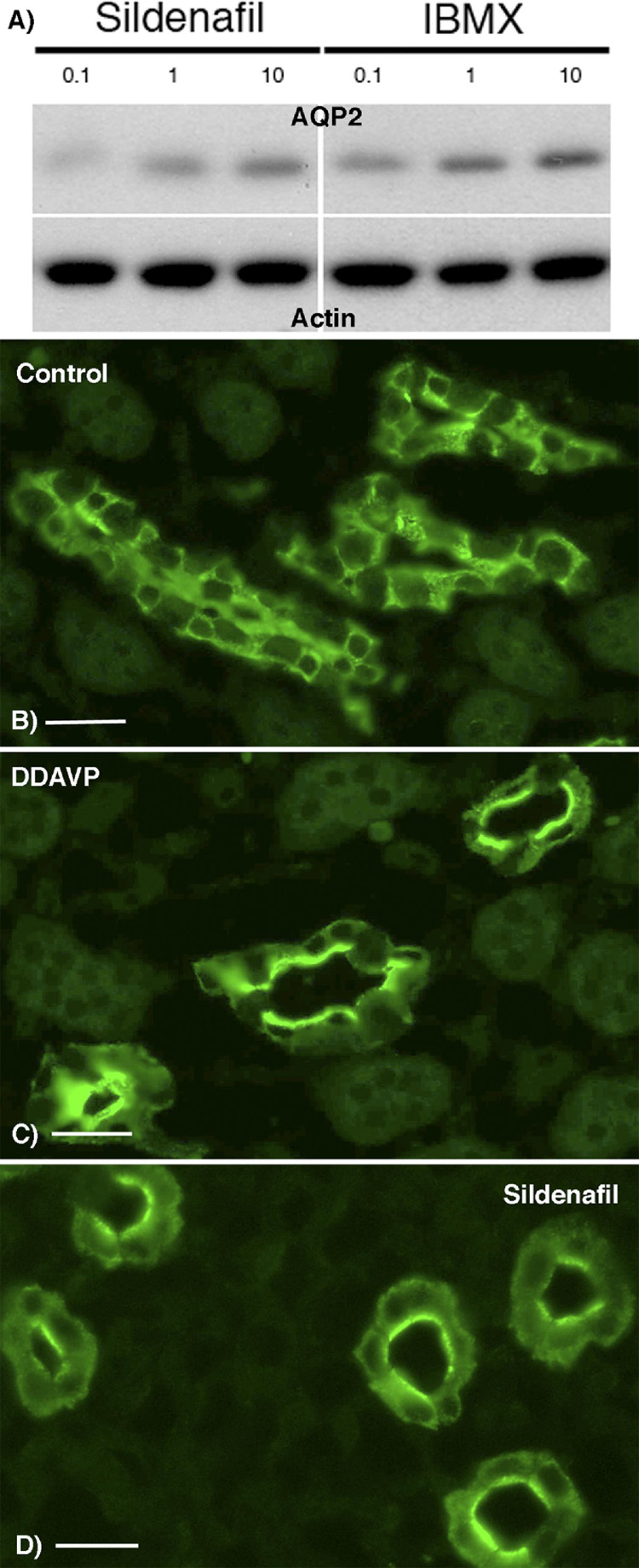
In panel A, cells were incubated 45 min in the presence of the selective PDE5 inhibitor (sildenafil) or with the non-selective PDE inhibitor (IBMX) at a 0.1, 1 or 10 fold higher dose than that corresponding to the EC50 of either chemical agent. A plasma membrane fraction was isolated from the cells and probed with anti-AQP2 antibodies. The same plot was reprobed with an anti-pan-actin monoclonal antibody as a loading control. Both sildenafil and IBMX induce the appearance of AQP2 in the plasma membrane fraction of the cells in a does-dependent manner.
In panel B kidney slices from a Sprague-Dawley rat were incubated for 10 min with (Deamino-Cys1, D-arg8)VP (DDAVP, 10 nM) or 45 min with sildenafil (0.5 µM) before fixation by immersion, sectioning and immunostaining for AQP2. Panel (A) shows a diffuse intracellular distribution of AQP2 in a control medullary collecting duct, whereas apical membrane accumulation (arrows) is induced in tissues treated with DDAVP (B) or sildenafil (C). Bar = 25 µm