
Figure 3. AQP2 membrane accumulation can be induced by inhibiting endocytosis.
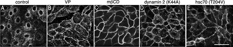
Control LLC-PK1 cells expressing AQP2 displayed baseline perinuclear AQP2 staining (A), whereas cells exposed to vasopressin (VP) showed strong AQP2 expression at the plasma membrane (B). Endocytosis was blocked in LLC-PK1 cells by methyl-β-cyclodextrin (mβCD) treatment (C), expressing a dominant interfering dynamin mutant (dynamin 2 DK44A) (D) or an ATPase deficient hsc70 mutant (T204V) (E). All three approaches to reduce endocytosis resulted in a dramatic increase of AQP2 expression at the plasma membrane. Immunostaining was performed using an anti-c-myc antibody to detect the c-myc tag of AQP2 in stably transfected LLC-PK1 cells. Bar = 20 µm.