
Figure 4. VP/FK treatment increases exocytosis in AQP2-expressing cells, but not in control cells.
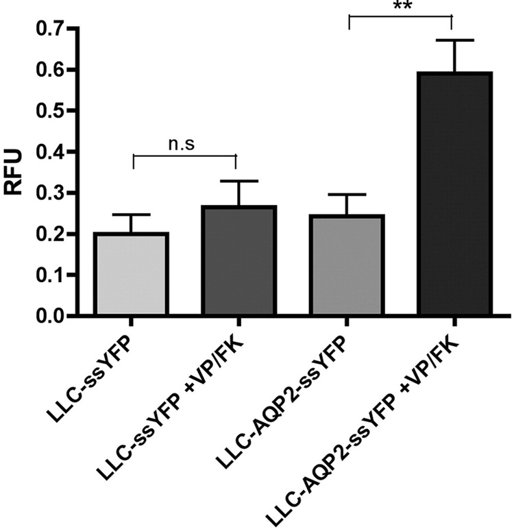
LLC-PK1 cells expressing AQP2 were transfected with a vector encoding a soluble, secreted form of yellow fluorescence protein, YFP (kindly provided by Jennifer Lippincott-Schwartz, NIH). The amount of ssYFP produced in LLC-ssYFP (which express YFP but not AQP2) and LLC-AQP2-ssYFP cells (which express AQP2 and YFP) and secreted in the extracellular medium after 15 min was measured by fluorimetry, and is similar between both cell lines under baseline conditions (bars 1 and 3 from left to right). When VP/FK is applied, AQP2-expressing cells show a large increase in ssYFP secretion within the first 15 min of stimulation, as compared to control cells (bars 2 and 4, respectively). These results are consistent with a large burst of exocytosis of AQP2-containing vesicles in response to VP/FK stimulation. Values were calculated as the relative increase from the 0 min baseline control and are expressed in relative fluorescence units (RFU). Each bar represents the average of 5 independent experiments performed in triplicate.